Back Muscle Diagram - Fill In The Blank Full Body Muscle Diagram - Voyeur Rooms / See back muscles and low back pain.. Both the deltoid and the trapezius are firmly attached to the spine of the scapula. To learn more about the anatomy of the spine, watch this video. Back muscles, like any other muscle in the body, require adequate exercise to maintain strength and tone. A clip from 3d back muscles: The back consists of the spine, spinal cord, muscles, ligaments, and nerves.
A video describing the major muscles of the back. There are several different layers of muscles in your back that are often pulling in different and various directions. Diagram of neck and back muscles diagram of neck and back muscles diagram of neck and back muscles upper back muscles cephalicvein. Muscles of the lower back and buttocks diagram, human muscles, muscles of the lower back and buttocks diagram. Barbell deadlift (from the floor)
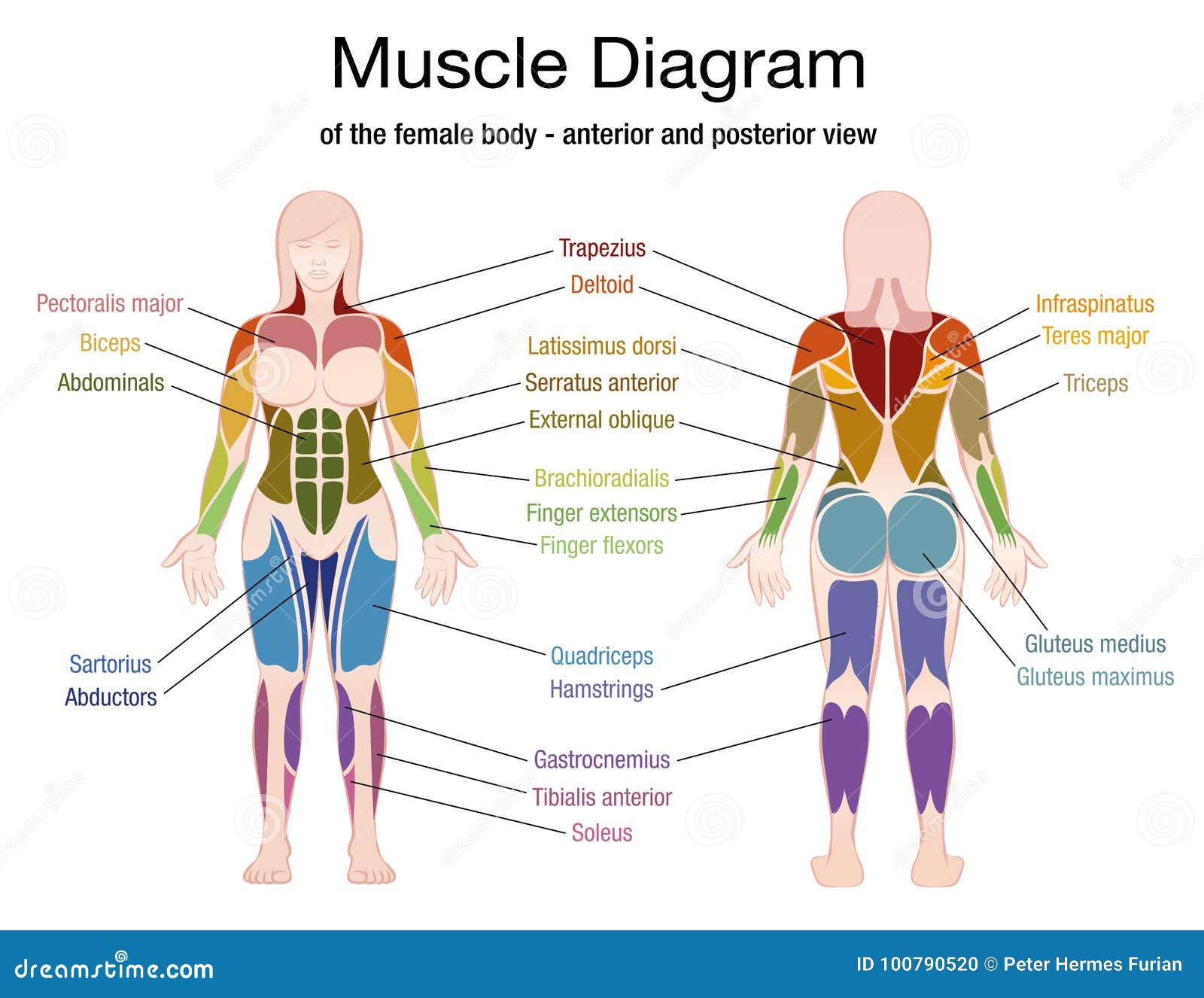
Muscle Diagram Female Body Names Stock Vector ... from thumbs.dreamstime.com Anatomy of the spine and back spine muscles diagram. The superficial back muscles are the muscles found just under the skin. However, the spinal erectors travel the length of the entire spine. Five pairs of lumbar spinal nerves labeled l1 to l5 branch off your spinal cord and exit through small holes between the vertebrae. The deep back muscles, also called intrinsic or true back muscles, consist of four layers of muscles: This can happen due to rigorous exercising, lifting heavy weights, or moving heavy items repetitively. The human back extends from the buttocks to the posterior portion of the neck and shoulders. Another common cause of lower back and hip pain is disc injury.
Diagrams of back muscles | 101 diagrams from www.101diagrams.com back muscle diagrams printable diagram.
A back muscle tear or injury similarly can be caused by overuse or strain of the muscles of the back. Back muscles, back muscle diagram. In this image, you will find 1st cervical vertebrae, atlus, cervical plexus, 7th cervical vertebrae, 1st thoracic vertebrae, brachial plexus, spinal dura mater, filaments of spinal nerve roots, 12th thoracic vertebra, 1st lumber vertebra, iliohypogastric nerve, ilioinguinal nerve, lumbar. The following diagram below is the diagrams of back muscle. The deltoid, teres major, teres minor, infraspinatus, supraspinatus (not shown) and subscapularis muscles (not shown) all extend from the scapula to the humerus and act on the shoulder joint. The most common type of back pain is muscle pain—also called muscle strain or soft tissue strain. Postural and active movement muscle, used to tilt and turn the head and neck, shrug, steady the shoulders, and twist the arms. See how exercise helps the back. This can happen due to rigorous exercising, lifting heavy weights, or moving heavy items repetitively. By the way, have you heard about the myth of. Back muscles, like any other muscle in the body, require adequate exercise to maintain strength and tone. What is the origin and insertion of the rhomboid minor and major muscle? Anatomynote.com found anatomy of back muscles diagram from plenty of anatomical pictures on the internet.
Diagrams of back muscles | 101 diagrams from www.101diagrams.com back muscle diagrams printable diagram. On these diagrams of back muscle, you'll learn about back muscles, their locations and functional anatomy. While muscles like the gluteals (in the thighs) are used any time we walk or climb a step, deep back muscles and abdominal muscles are usually not actively engaged during everyday activity. Both the deltoid and the trapezius are firmly attached to the spine of the scapula. We hope this picture anatomy of back muscles diagram can help you study and research.
Muscle Model Labeled - Top Label Maker from labels-top.com Deadlift variations for back growth: A video describing the major muscles of the back. The deep muscles develop embryologically in the back, and are thus described as intrinsic muscles. Muscles of the lower back and buttocks diagram, human muscles, muscles of the lower back and buttocks diagram. Superficial back muscles, intermediate back muscles and intrinsic back muscles.the intrinsic muscles are named as such because their embryological development begins in the back, oppose to the superficial and intermediate back muscles which develop elsewhere and are therefore classed as extrinsic muscles. Nerves in your lower back. Most of the time, back muscle pain is diagnosed then treated with little more than a prescription of rest, painkillers and muscle relaxants. The latissimus dorsi, also known as the lats or wings, are.
Muscle spasms (contraction or stiffening of the back muscles) muscles that feel tight;
To learn more about the anatomy of the spine, watch this video. The human back extends from the buttocks to the posterior portion of the neck and shoulders. Superficial, intermediate, deep and deepest layers. How many muscles are in the back? The most common type of back pain is muscle pain—also called muscle strain or soft tissue strain. Creatine is now proving to be one of the most potent muscle growth accelerators giving excellent muscle mass increase and phenomenal strength increases order yours today. These muscles lie on each side of the vertebral column , deep to the thoracolumbar fascia . The superficial back muscles are the muscles found just under the skin. It is opposite from the chest, and the vertebral column runs down the back. It comprises the vertebral column (spine) and two compartments of back muscles; A clip from 3d back muscles: Superficial back muscles, intermediate back muscles and intrinsic back muscles.the intrinsic muscles are named as such because their embryological development begins in the back, oppose to the superficial and intermediate back muscles which develop elsewhere and are therefore classed as extrinsic muscles. In this image, you will find 1st cervical vertebrae, atlus, cervical plexus, 7th cervical vertebrae, 1st thoracic vertebrae, brachial plexus, spinal dura mater, filaments of spinal nerve roots, 12th thoracic vertebra, 1st lumber vertebra, iliohypogastric nerve, ilioinguinal nerve, lumbar.
We hope this picture anatomy of back muscles diagram can help you study and research. The human back extends from the buttocks to the posterior portion of the neck and shoulders. What is the origin and insertion of the rhomboid minor and major muscle? Five pairs of lumbar spinal nerves labeled l1 to l5 branch off your spinal cord and exit through small holes between the vertebrae. For more anatomy content please follow us and visit our website:
Muscular System Worksheet | Lobo Black | Muscle diagram ... from i.pinimg.com The muscle elevates, depresses, rotates, and retracts the scapula, or shoulder blade. However, the spinal erectors travel the length of the entire spine. Back muscles, back muscle diagram. While muscles like the gluteals (in the thighs) are used any time we walk or climb a step, deep back muscles and abdominal muscles are usually not actively engaged during everyday activity. Barbell deadlift (from the floor) Some of the links in the post above are affiliate links.. Muscles of the back diagram. What is the origin and insertion of the rhomboid minor and major muscle?
See how exercise helps the back.
Know the causes, symptoms, treatment, recovery period of back muscle tear or injury. Five pairs of lumbar spinal nerves labeled l1 to l5 branch off your spinal cord and exit through small holes between the vertebrae. Back muscles, like any other muscle in the body, require adequate exercise to maintain strength and tone. Back muscles chart, back muscles diagram and ligaments, back muscles diagram lats, back muscles diagram massage, upper back muscles chart, human muscles, back muscles. These structures work together to support the body, enable a range of movements, and send messages from the. However, the spinal erectors travel the length of the entire spine. Both the deltoid and the trapezius are firmly attached to the spine of the scapula. Daniel nelson on january 1, 2019 2 comments 🔥! Lower back muscle and hip pain may also be caused by stenosis in the spine. The muscles of your back support your spine, attach your pelvis and shoulders to your trunk, and provide mobility and stability to your trunk and spine. The most common type of back pain is muscle pain—also called muscle strain or soft tissue strain. Creatine is now proving to be one of the most potent muscle growth accelerators giving excellent muscle mass increase and phenomenal strength increases order yours today. The muscles of the back can be arranged into 3 categories based on their location: